Stage One: Preparation of kinase
1. Preparation of Kinase in Solution: Prepare the purifiedor partially purified (fractionated) kinase SGK in 10 µL of the kinase assay dilution buffer (component D) with the required concentration in microcentrifuge tubes. For inhibitorscreening, serial dilutions (range from 1000 to 50ng/assay) of kinase should be tested for the optimal working concentration.
2. Preparation of the Immunoprecipitated Kinase:
Incubate anti-SGK (able to IP native kinase), or anti-GST, anti-HA (for tag-kinase) with 10 to 20 µL of protein A for 1 hr. Then wash away any unbound antibodies. Incubate the cell lysate in 250 µL of lysate buffer with the antibody-protein A complex in a rotor shaker at 4oC for 2 hrs. The user should select their own cell lysate buffer that contains protease and phosphatase inhibitors. The cell lysate sample should contain >20 ng of active SGK. Centrifuge and aspirate with PBSt twice then with kinase assay dilution buffer (D) twice. After aspiration, re-constitute the immunoprecipitated matrix in 10 µL of kinase assay dilution buffer (D).
3. Preparation of Kinase Purified with Affinity Matrix (GSH, Ni, maltose, etc.): The user should purify therecombinant kinases, such as GST-SGK, following their own protocol. 10-20 µL of affinity matrix/assay is recommended. Do a final wash and aspiration of the matrix with kinase assay dilution buffer (D) then re-constitute the aspired matrix with 10 µL of the kinase assay dilution buffer.
Stage Two: SGK kinase activity assay
1. Add 2.5-5 µL of the BSA-kinase substrate (component C) to the kinase sample prepared in stage one.
2. Add 5 µL of the ATP (component E) solution to initiate the kinase reaction. Incubate at 37oC for 60 min with constant shaking or hand shake every 5 min.
3. Stop the kinase reaction with 20 µL of SDS-sample loading buffer and boil for 2 min.
Stage Three: Running SDS-PAGE and transfer;
1. Prepare a 8% SDS-acryamide gel.
2. Run the SDS-PAGE until the dye-front is approximately 2-3 cm past entering the separation gel.
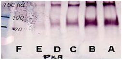
3. Perform the normal gel transfer to nitrocellulose membrane. The membrane only needs to cover between the top of separating gel and the distance to MW marker 60 kDa.
Stage Four: Immunoblotting.
1. Block the membrane with 3% BSA or 3% skimmed milk for 30-60 min
2. Wash the membrane twice with PBSt.
3. Incubate with 10 mL of anti-pSubstrate (component A), 0.25-0.5 µg/mL in antibody dilution buffer (component B) at room temperature for 2-4 hours, or 4oC overnight.
4. Wash the membrane with PBSt 4 times at 5 min intervals.
5. Incubate with anti-rabbit IgG secondary antibodies for 1-2 hours.
6. The user should select their own method (either colormetric or ECL system) for the signal generation.
